新入荷
再入荷
【おしゃれ】 Atlas of Pathologic Myopia 【英語版】 - 本 健康・医学
 タイムセール
終了まで
00
00
00
999円以上お買上げで送料無料(※)
999円以上お買上げで代引き手数料無料
999円以上お買上げで代引き手数料無料
通販と店舗では販売価格や税表示が異なる場合がございます。また店頭ではすでに品切れの場合もございます。予めご了承ください。
商品詳細情報
| 管理番号 |
新品 :69744130329
中古 :69744130329-1 |
メーカー | 844a221d090e25 | 発売日 | 2025-07-14 14:58 | 定価 | 15000円 | ||
|---|---|---|---|---|---|---|---|---|---|
| カテゴリ | |||||||||
【おしゃれ】 Atlas of Pathologic Myopia 【英語版】 - 本 健康・医学
 Atlas of Pathologic Myopia 【英語版】 - 本,
 Advances in OCT Imaging in Myopia and Pathologic Myopia,
 Pathologic myopia: advances in imaging and the potential,
 Advances in OCT Imaging in Myopia and Pathologic Myopia,
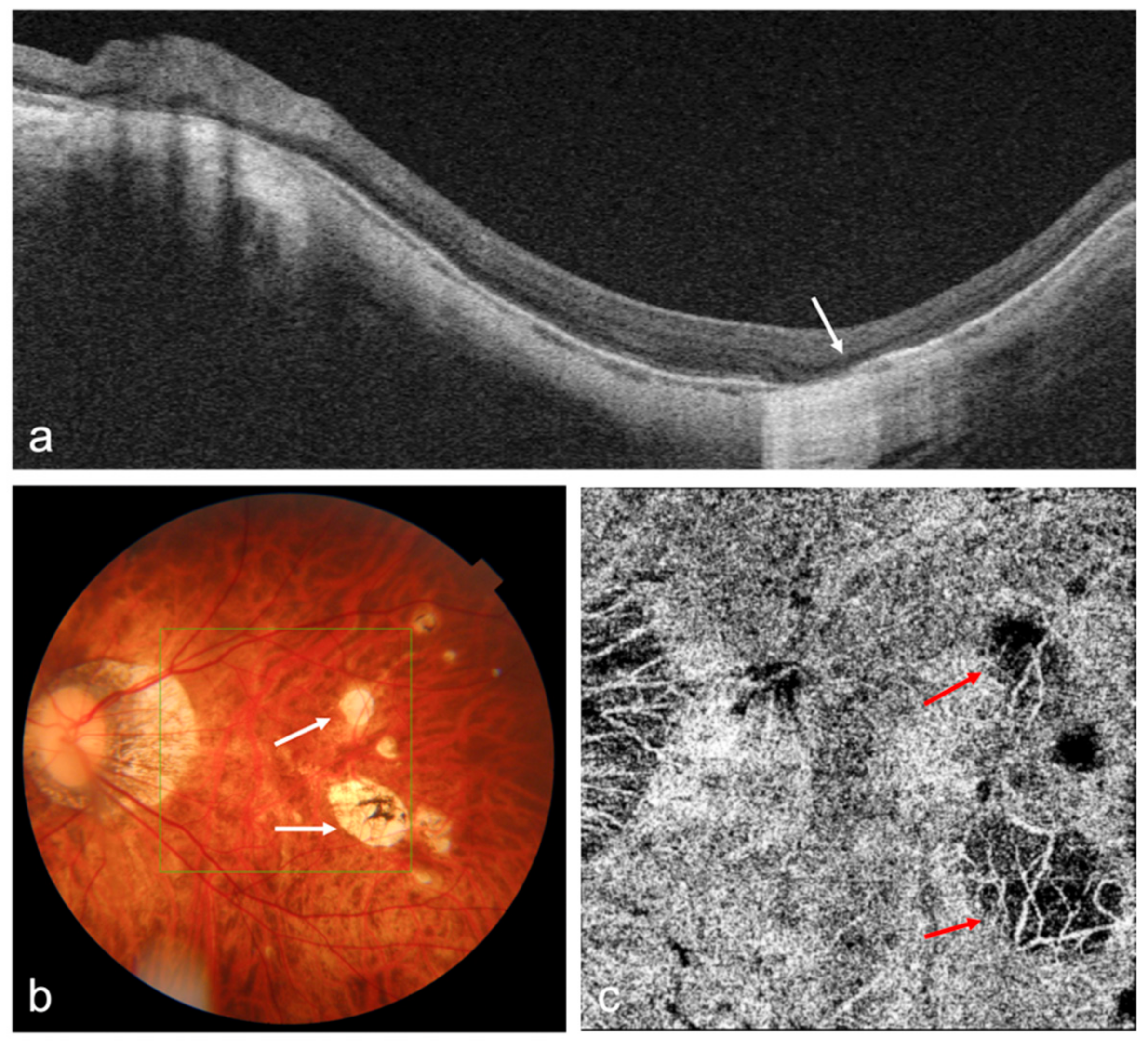
 Frontiers | Global trends and frontiers of research on【ハードカバー】の書籍です。第62回麻酔科専門医認定試験対策資料。\r編者:Kyoko Ohno-Matsui\r出版社:Springer\r2020版 114ページ\r\r一般サイトで¥27000程です。グレイ解剖学 原著第4版。\r\r※プロフィールをご一読お願いします。糖尿病専門医研修ガイドブック 改訂第9版。\r※学生の方は申し出てください\r\r使用の見込みがないため出品します。医学書セット X線・MRI・CT関連。\r実際は未使用ですが、少々のスレが感じられるので、未使用に近いとしました。口腔外科 教科書。表紙、中身はかなり綺麗です。【専用】午前中希望 プロフ必読様。\r\r梱包封筒で発送します。DVD>神経系モビライゼーション 上肢編。非喫煙者、ペットなしです。《裁断済み》最強のクリニック経営術※My※。\r\r内容:\r世界最大の高度近視クリニックが提供する豊富な画像を特徴とする 最先端技術による画像 多数の症例シリーズの治療適応とアウトカムを収載\r\rThis Atlas provides many beautiful images obtained with state-of-the-art technologies, including optical coherence tomography (OCT), OCT angiography, fundus autofluorescence, and wide-field fundus imaging, as well as traditional images and fluorescein/ICG angiograms. Gathered at the world's largest High Myopia Clinic, the images are based on the long-term follow-up data of more than 6,000 patients from Japan and abroad. Recent advances in imaging technologies have yielded many new observations and allowed us to detect new lesions, e.g. myopic traction maculopathy (or macular retinoschisis) and dome-shaped macula. An especially interesting aspect: the images obtained by `3D MRI of the eye..


